Leg veins come in many different sizes, colours and patterns and there are a range of treatment options. Recognising the type of leg veins you have can help in understanding which treatment will work best.
Leg Veins fall into 3 main types based on size:
- Spider Veins: Less than 1 mm diameter
- Reticular Veins: 1-4 mm in diameter
- Varicose Veins: Larger than 4 mm diameter
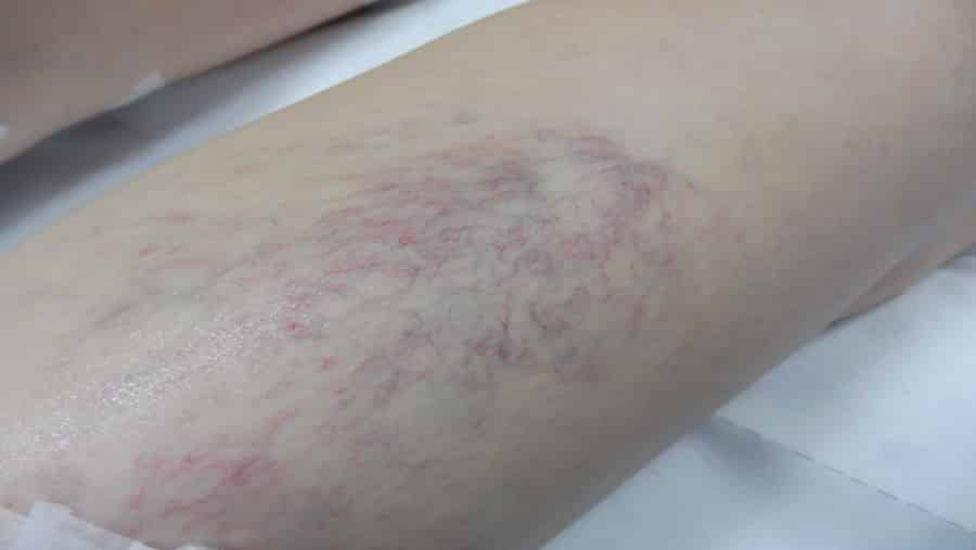
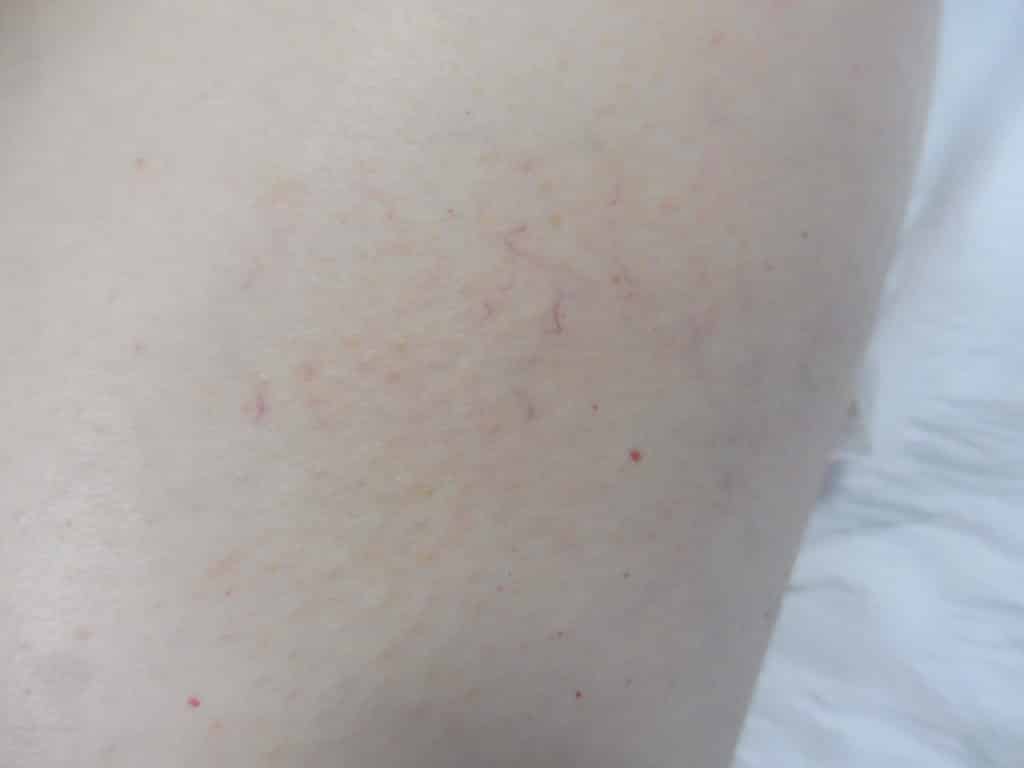
Spider Veins are so called because they resemble the strands of a spider web. Medically we refer to Spider Veins as Telangiectasia and the colour can vary from bright red for the smallest Spider Veins through to dark blue for larger Spider Veins. Spider Veins appear in various patterns such as scattered, in clusters or as thick patches that can resemble a bruise (called a Venular Flare). Spider Veins can occur in isolation or in combination with larger Reticular or Varicose Veins in the same area.
Treatment of Spider Veins is by a surface treatment either microinjections (Surface Sclerotherapy), Laser or the Veingogh (Surface Radiofrequency). The smallest bright red Spider Veins respond well to Surface Laser or Veingogh whilst larger Spider Veins usually respond faster and achieve better results with Sclerotherapy. Before treating Spider Veins it is important to make sure there are no deeper underlying vein problems as these also may need to be treated. When there is a combination of Spider Veins and Varicose Veins typically the largest veins are treated first.
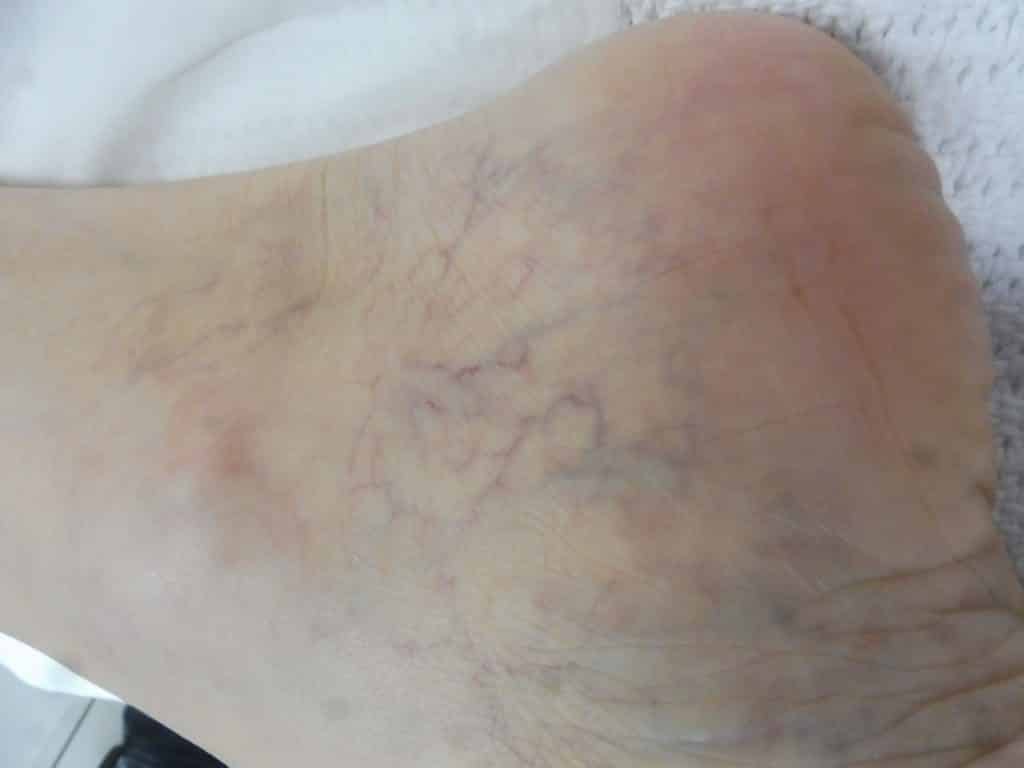
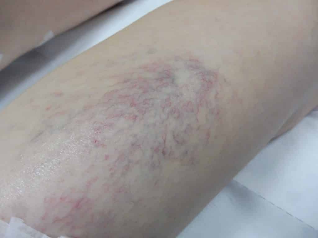
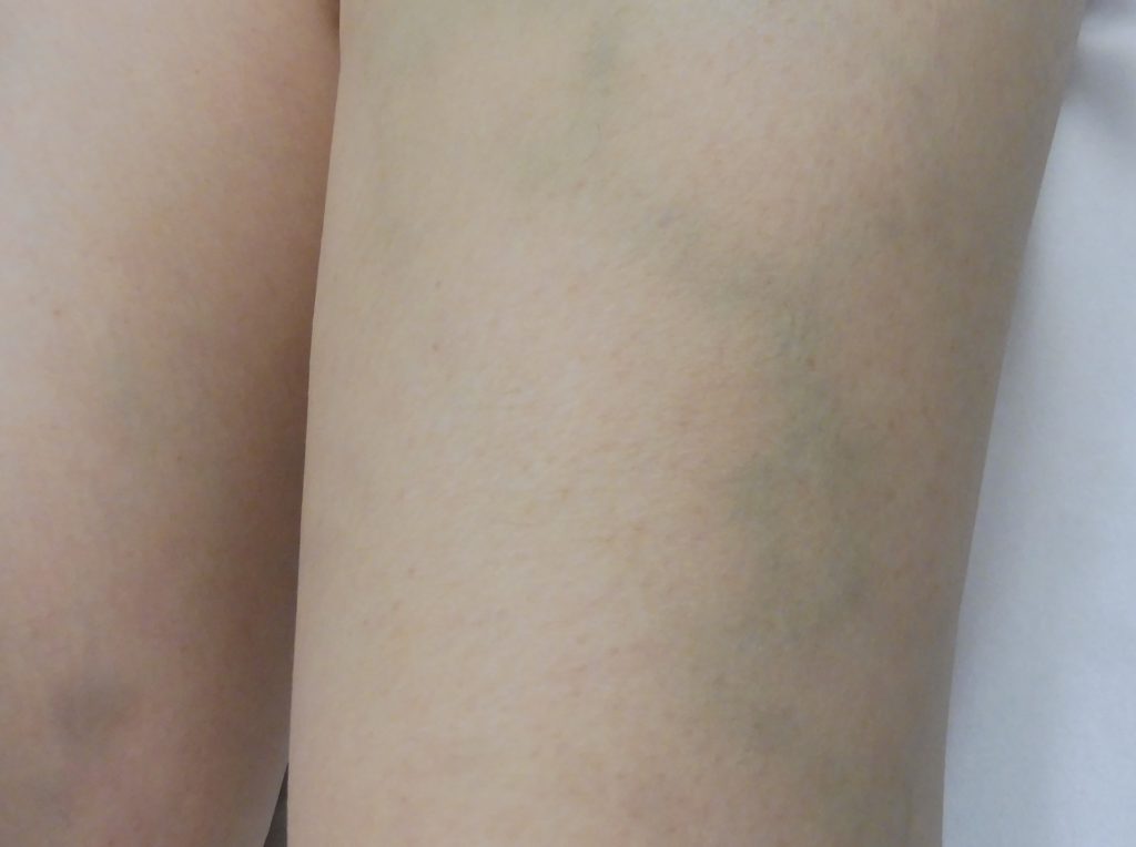
Reticular Veins are larger than Spider Veins but smaller than Varicose Veins and are blue in colour. The best treatment option for Reticular Veins is Sclerotherapy. The treatment of Reticular Veins often requires Foam Sclerotherapy which uses a thicker solution to shut down the larger veins more effectively than the standard liquid Sclerotherapy.
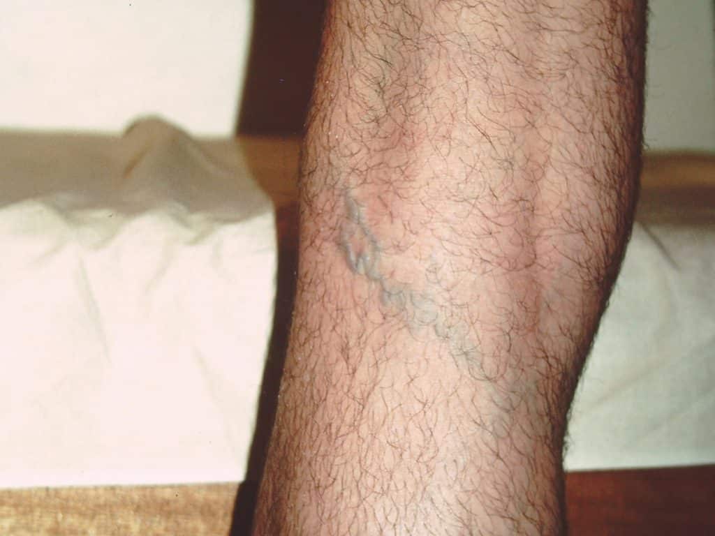
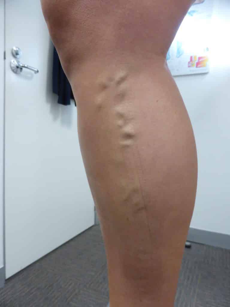
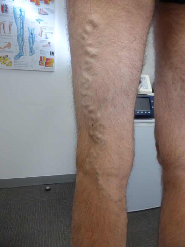
Varicose Veins are the largest diameter veins. The word Varicose means enlarged, dilated and tortuous and most people regard Varicose Veins as those veins that bulge above the surface of the skin.
There are several ways to classify Varicose Veins and whilst they are regarded as any veins larger than 4 mm in diameter the most important distinction in deciding the best treatment option is if
the Varicose Vein originates from the main deep vein (called a Truncal Varicose Vein) or from a branch of the main vein (called a Branch Varicose Vein). This distinction requires Ultrasound scanning and for the best long term results in treating Varicose Veins we need to treat where the problem is coming from not just the Veins that are visible on the surface of the skin.
